What happens during a Heart Attack?
MRI – Non-Invasive Window into the Heart
Doctors use magnetic resonance imaging (MRI) as a non-invasive, diagnostic tool to look at the soft tissue inside of a patient without having to invade the body. MRIs use a powerful, but harmless magnetic field that reveals in great detail the shape and condition of your internal organs. Doctors can use this test to diagnose various conditions of the heart and the entire body.
What can this non-invasive MRI procedure tell a doctor about a patient’s heart and circulatory system? Miracle MRIs can identify heart scarring and any other indications of a previous or future heart attack. It can reveal arterial clogging and the presence of other foreign masses in, and around, the heart and body. It detects any signs of heart disease, identifies vascular disorders, and checks blood vessel health. Warning: MRIs cannot be used on people with pacemakers or arterial clips.
Safer and Healthier Non-Invasive Tests For Heart Disease
DOPPLER COLOR FLOW IMAGING TEST
Non-invasive imaging ultrasound. It shows a clear profile that checks the entire blood vessel system simultaneously. When needed, doctors check for possible blood slowdown and blockages that can cause future heart attacks and strokes, and even death.
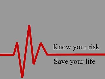
ELECTROCARDIOGRAM & CARDIOKYMOGRAPHY
It’s an EKG and CKG graph that records electrical activity in the heart. The heart has an electric current running through it. The heart contracts and pumps blood throughout your body. This contractionis started off by an electric current, even though it is a weak one. This current begins in a part of the heart called the sino-atrial node, or the pacemaker, which sets the pace for the heart to beat. From the pacemaker this current follows a well-defined path through the rest of the heart. This movement can be recorded by electrodes, which are plastic plates placed on the chest and limbs to detect the current flow inside the heart. The graph recorded is the EKG and CKG, both painless tests. These tests detect disturbances in the pattern of electrical activity in the heart, called arrhythmia. They also check if any chamber of the heart is abnormally enlarged or if any of the walls have thickened.
ECHOCARDIOGRAPHY
This painless ultrasound is an examination of the heart, used to evaluate structural conditions like the thickness of the walls; the way the heart walls move during exercise or rest; diagnose valve trouble; inflammation; congenital heart disease and congestive heart failure. Echocardiography uses high-frequency sound waves to produce images of the heart. A small transducer, like a microphone, passes over the chest, sending out impulses that bounce off the heart. The transducer records these echoes, and a computer converts them into a graphic display on the screen.
EXERCISE STRESS TESTING
This is an exercise EKG electrocardiogram that’s performed with controlled exercise such as a treadmill. The patient’s maximum heart rate is calculated based on their sex and age, and then the patient is connected to the EKG machine and exercises until the heart is beating steadily at the calculated rate. This test shows changes in the EKG pattern, especially for those with narrowing of the coronary arteries. If blood pressure drops during the test, this could be another sign of coronary artery disease. The stress test is also used for people who recently recovered from a heart attack, as an initial step in assessing the heart’s blood supply. Please express any sensations experienced during testing (sometimes it’s too much too soon). This test can detect coronary artery disease in 75% of cases.
NUCLEAR SCANNING
This safe technique uses radioactive materials known as isotopes, to examine the heart. The isotopes used are harmless substances, and less radioactive than most x-rays. In nuclear scanning, the patient is given a small dose, either orally or injected. These isotopes flow through the blood system giving off radiation which is photographed by a special camera producing pictures of the heart. These pictures show how well the ventricles are working and where there is scarring, damaged or oxygen-starved areas of the heart.
HOLTER MONITOR
Is a portable version of an EKG. It records the heart rhythm (pulse) during daily activities. This is worn for 24 hours or more. The heart waves are picked up by electrodes or patches placed on your chest. These waves are recorded on a tape inside the monitor. This recording is then scanned into a computer for analysis. Holter Monitors are used on patients who experience chest pain, dizziness, palpitations, or fainting, most often caused by narrowing of arteries or heartbeat abnormalities. It may also show evidence of silent ischemia, which is like an angina attack (page 16) without chest pain.
New Millennium Health Technology Provides Safer, Faster, Better Testing
DIGITAL TECHNOLOGY
Can take a routine test like a chest x-ray to a new height of quality and has many advantages. It’s an environment saver because there are no film processing chemicals utilized; it is safer for the patient since it uses lower x-ray doses and reduces need for retakes and more exposure; it gives an instant picture and can be stored on a computer and transmitted instantaneously to the doctor’s office, hospital or anywhere!
PET SCANNER: Positron Emission Tomography
Is a unique 32-ring scanner that can detect and measure metabolic activity throughout the body and especially the brain It pinpoints the source of cancers, neurological and heart diseases; thereby reducing all the expensive, unneeded operations, exploratory surgery and hospital stays! The PET scanner saves time, money and most important, precious lives!
FOR SURGERY: XKNIFE STEROTACTIC RADIOSURGERY
Uses a radionic invisible blade not a scalpel; this makes surgery non-invasive, bloodless, reduces complications, discomfort, hastens recovery. Excellent for brain tumors and arteriovenous malformations (AVMs). (www.radionics.com)
When needed, seek the highest quality, safest testing technology. Use these valuable guidelines as a source. It’s better to be safe than sorry. Medical and Hospital Emergency Centers do fast tests to relieve your mind to see if you have had a mild heart attack.
Dr. Paul Dudley White of Boston – Famous Heart Specialist’s Wise Words
Dr. White,the former president of the American Heart Association, world famous pioneer heart specialist and our friend, gave this wise advice on taking care of the heart. We want to call your attention especially to the following points made by Dr. White in an article written for the American Heart Association. He begins with his startling facts that middle age begins at 20, and the dangerous years are ages 20 to 40! Here are Dr. White’s words:
When does middle age begin? At 20, and it lasts until 80. And the dangerous years of this span are the first 20, not the last. These are the years when an overfed and under-exercised public is sowing the seeds of a coronary harvest.
I conceive the ages of man as five, Dr. White continues. Birth to the 20th year; then a three stage middle age of 20 to 40, 40 to 60, 60 to 80; and finally old age – 80 to 100. The latter constitutes a steadily expanding horizon to which I see no eventual limit. Our life expectancy should keep rising indefinitely as research keeps making progress against disease.
Unlimited Life Expectancy is Possible!
Dr. White stated, the public can play an important role in this effort to push the life-span farther and farther. Physical-fitness and nutritional programs for men and women between the ages of 24 and 40 would guard against creeping degeneration and would instill lifelong good health habits.
A man marries in his 20s; his wife cooks too much and too well – and between her cooking, the family car and the TV set, the man has gained maybe 20 to 30 pounds by the time he’s 45. These are the years in which atherosclerosis (cholesterol blocking and clogging the arteries) and rusting of the arteries occur. This can ultimately reach the brain as a stroke, or the heart as a coronary thrombosis (massive blood clot). It may also affect the kidneys. This is why an apparently healthy man drops dead at 45 or 50. His death is not sudden at all; an unhealthy lifestyle has silently been building up for years!
Thank you – your explanation of the homocysteine theory of heart disease made this a red letter day in my life! – Dr. Paul Dudley White to Dr. Kilmer S. McCully, author The Homocysteine Revolution.
The automobile and the TV, I might add, should be the servants of the American public, not its masters. Despite the nation’s generally unhealthy way of life, two factors work in favor of the American person, Dr. White concludes. It is never too late – at any age – to begin controlling obesity and resuming a program of sensible exercise and a healthy diet. One excellent form, available to all, is walking. This should be brisk, and for a normally healthy person five miles weekly is not enough. Neither is one weekly 18-hole golf game.
There you have it – from Dr. White, known as Dean of American Cardiology. Exercise and diet can be regular and enjoyable parts of the Healthy Lifestyle.


