Cholera: Causes, Symptoms, Treatment, and Prevention
CHOLERA – WORLD HEALTH ORGANIZATION
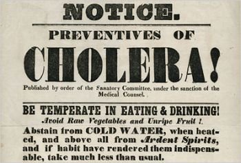
Cholera
A bacterium that is equally swift at killing is Vibrio cholerae, the causative bacterium for cholera. This water-borne bacterium has also been responsible for pandemics, seven in the last 200 years according to the World Health Organization. Because of the low speed of transport and the rapid onset of symptoms, before the mid 19th century, cholera outbreaks were largely contained in and around the country in which they emerged. The first pandemic originated in the Indian subcontinent or China, in common with those pandemics that followed. Cholera was endemic in the region surrounding the Ganges, and pilgrims to the festivals carried the organism back to their own villages and towns. This had happened in previous outbreaks but, by the 1820s, British soldiers carried it to areas far from the subcontinent including Africa and the rest of Asia.
The second pandemic spread more quickly during the 1830s as communications had improved. Again starting in India, it spread to Europe, Russia, Canada, and the United States. It was responsible for a number of deaths in the British Isles up to the end of the 1840s. However, the cause of the infection was not understood until the third pandemic reached London in 1853 and John Snow identified that contamination of the water supply was responsible. Even so, this pandemic was responsible for more than 10,000 deaths in London; however, it was the last time that cholera seriously affected the United Kingdom. Further pandemics occurred during the 19th century but better hygiene largely prevented cholera seriously affecting Europe, except for the fifth pandemic, causing a major outbreak in Hamburg in 1892, and some parts of Eastern Europe affected by the sixth pandemic in the early 20th century.
We are still experiencing the final and seventh pandemic. It started in the 1960s, this time in Indonesia. The causative bacterium was not the classic strain but a related strain called El Tor. It had originally been isolated from a cholera patient being treated in El Tor in Egypt in 1905. The strain spread from Indonesia to the subcontinent and North Africa. From North Africa it spread to Italy, though the number of cases was relatively few. This strain spread to South America, in particular Peru and northern Chile, and from there to the southern United States. El Tor is a milder pathogen than the classic strain and its symptoms are less severe; however, the main controls of the current pandemic have been better hygiene, preventing infection in the first place, and much more effective treatments, reducing mortality.
Leprosy and tuberculosis
Plague and cholera are infections of rapid onset and swift mortality. It is thus easy to follow their spread across the world. Two related bacteria are far slower in their transmission; they are Mycobacterium leprae and Mycobacterium tuberculosis, the causative bacteria for leprosy and tuberculosis. For agar in a Petri dish t0R this reason they have been much more difficult to control.
Leprosy and plague were considered to be the two major epidemics of the Middle Ages and leprosy remained so until the 18th century. Certainly leprosy was common in Europe, though it did not follow classic pandemic distribution. Indeed, in many areas it was probably endemic. Leprosy is an infection of the skin and nerves; although it is contagious, a person has to have long exposure before the bacterium can establish an infection. There has been a progressive decrease in the number of infections, partly due to better and more rapid diagnosis and subsequent antibiotic treatment but also possibly to less crowded living conditions and an improved host immune status. India still has the majority of cases, mostly in poor communities, but the disease is found in other countries of the subcontinent, parts of Africa, and in Brazil. Currently, the proportion of cases in the world is now only 10 per cent compared with the past so this is a bacterial pandemic that is also declining.
The same used to be true for tuberculosis as it was for most of the major bacterial infections but now the number of cases is increasing. Also slow growing, Mycobacterium tuberculosis can spread more quickly as it is usually airborne and coughing is a usual symptom. The universality of air travel is thought to promote the spread of tuberculosis but there is no strong evidence for this. Probably the major cause of the resurgence of tuberculosis is the increase in infections by Human Immunodeficiency Virus (HIV), which undermines the body’s defences against tuberculosis so that it is a common accompanying infection. Tuberculosis is a disease particularly of the urban poor, not just in the developing world but also in the ghettos of industrialized countries, with inadequate access to healthcare being a further contributing factor.
Infection can only occur from those suffering from active infection and is transmitted on droplets. The infective dose of Mycobacterium tuberculosis is as low as 10 bacterial cells, so any droplet, in theory, could initiate an infection. Prolonged contact increases the risk, as does the virulence of the strain and how well the environment is ventilated. The vast majority of those infected do not develop the disease but it may remain latent within those infected. The risk of progression to active tuberculosis infection depends on immune status, due to either infection or previous history of transplant requiring immunosuppression, cancer and other underlying medical conditions. Low body mass is also associated with active infection, which probably accounts for its increased transmission in deprived communities and, partly for these reasons, the epidemic has been difficult to control. Furthermore, a major risk factor is diabetes mellitus, and the number of these cases is currently increasing worldwide. The extent of the tuberculosis epidemic at the start of the 20th century was a major incentive to find drugs that could cure it. However, the ‘White Death’ was beginning to decline in the United Kingdom.
Living conditions improved and at some point around 1935, the number of rooms in a family dwelling exceeded the number within the family. This had a considerable influence in reducing the spread of all types of infection, not just tuberculosis. It was, however, the discovery of Graph showing the decline of bacterial infections over the last century ret streptomycin, one of the earliest antibiotics, that broke the tuberculosis epidemic cycle in the United Kingdom, and appropriate timely treatment can provide a cure. In many areas, poor medical supervision and insufficient dosing have resulted in a resurgence of antibiotic-resistant tuberculosis. Treatment of tuberculosis requires daily administration of antibiotics for as long as six months, almost impossible in some countries. There are very few options for infections caused by these bacteria. One method of control has been vaccination with the Bacillus Calmette-Guérin, commonly known as BCG, an attenuated live tuberculosis strain from cattle. It does provide some protection for about fifteen years but largely in those countries where tuberculosis is on the decline. The efficacy of BCG is considerably lower in countries where the bacterium is still responsible for an epidemic.
The extent of the epidemic has ensured that one-third of the world’s population has been infected. Though most are asymptomatic, in some countries the proportion of the population with active infections is around 2 per cent and it is compounded by the fact that 20 per cent of these strains are resistant to the main antibiotics. Until this situation is reversed, there is little prospect that the epidemic will dissipate.
Sexually transmitted infections
Currently, the most feared sexually transmitted disease is caused by a virus, Human Immunodeiciency Virus (HIV). Nearly 1 per cent of the world’s population is infected. In the United Kingdom, the figure is under 0.2 per cent but rises to 5 per cent in sub-Saharan Africa. In contrast, it has been suggested that at the turn of the 20th century, 10 per cent of the adult male population in the United Kingdom had syphilis. This bacterial epidemic, then untreatable with antibiotics, was as shocking as the HIV epidemic is today and had an eventual mortality rate of up to 50 per cent. The social taboos of the time led many men to have their early sexual experiences with women who had had many sexual partners, particularly prostitutes, who provided a reservoir for the epidemic. The bacterium, Treponema pallidum, is commonly believed to have to come to Europe in the 15th century from the Americas, though there is some evidence that it was prevalent in Ancient Greece and Rome. It spread rapidly during the Renaissance, facilitated by the migrations occurring at that time and, because of its symptoms producing pustules like smallpox, soon earned its nickname of ‘the Pox’. Syphilis was widely treated with mercury but it was probably due the promotion of condoms, particularly by the military in the late 19th and early 20th centuries, and the introduction of Salvarsan in 1912 that the incidence began to decrease. The advent of antibiotics reduced the epidemic so that by the end of the 20th century there were only sporadic cases in the developed world, though it is still prevalent outside Europe and North America.
Other major bacterial sexually transmitted diseases include chlamydia and gonorrhea, and these have not shown such a marked decline; they are still the two most common sexually transmitted bacterial infections. Gonorrhea, caused by the bacterium Neisseria gonorrhoeae, has been documented in England since at least the 12th century and has been presumptively diagnosed in men by a burning sensation while urinating. The problem with gonorrhea is Graph showing the decline of bacterial infections over the last century ret that a woman has a greater risk of becoming infected from an infected man than a man from an infected woman. As it is initially asymptomatic in many women, the infection can spread unnoticed, making it difficult to track. The work of the clinics that treat infected patients is not just the administration of antibiotic but also tracing previous sexual partners. Through this extraordinarily difficult and sensitive task, many contacts can be treated who have shown no symptoms. The incidence of gonorrhea fell with the introduction of condoms, antibiotics, and more recently fears about infection with HIV. Early treatment of gonorrhea soon clears the infection but, if left, it can cause serious medical problems.
Chlamydia is a more prevalent sexually transmitted disease. Like gonorrhea, most women show no symptoms when infected and the bacterium can remain undetected for years. In both sexes it can lead to inflammation in the genitals, but this often occurs long after the bacterium has been passed to other partners. The bacterium, Chlamydia trachomatis, only multiplies in human cells and can be difficult to detect. Indeed, it cannot be detected by culture but rather by modern DNA techniques. These factors ensure that chlamydia is a silent pandemic as many cases are not detected and current assessments of the infection rate are almost certainly underestimates.
Bacterial toxins as weapons
In the Middle Ages, during the siege of a town as the population was starving, contaminated meat or even faeces were often catapulted over the wall in an attempt to weaken the besieged population by disease. There were even attempts to introduce plague into these towns. The problem with this early form of germ warfare is that the aggressors could not control the extent of the infections and were often infected themselves once the town had been taken.
A bacterium was needed that could be contained and did not pose a threat to the army using it. Anthrax, caused by Bacillus anthracis, was a convenient choice. This spore-forming bacterium has a very high mortality rate, does not spread extensively from its target area and does not spread from person to person. The bacillus was isolated by Robert Koch in 1876 and attempts were made to use it during the First World War, particularly against livestock and the horses of the cavalry on both sides.
Antibiotics
It is likely that a few antimicrobials have been used by some indigenous populations for centuries. Quinine, from the bark of the cinchona tree, had been used by Indians in Peru, and was effective against malaria; this knowledge was brought back to Europe in the 17th century when it became possible to control malaria. However, there are more than a hundred prescription medicines that are derived from the plants of the rainforest, and some have antibacterial properties. As the indigenous populations became adept at exploiting these plants, they were able to control some bacterial infections.
Oliver Wendell Holmes published a paper in the New England Quarterly Journal of Medicine on the ‘The Contagiousness of Puerperal Fever’ in 1843. At the time, the role of bacteria, or any microorganism, as a cause of infection was unknown. Holmes concluded that puerperal fever, contracted by women during childbirth, came from person-to-person contact probably from the physicians treating them. He also noted that a number of physicians had died after performing autopsies on patients who had been infected. Holmes suggested that for physicians who had performed autopsies on patients who had died of puerperal fever, their surgical instruments should be sterilized and their clothing should be burnt. His most drastic conclusion was that the physician should not participate in childbirth for the following six months.
Ignaz Semmelweis, a Hungarian doctor working in Vienna in 1847, had a less drastic solution. He noticed that in those wards where the doctors washed their hands after they had performed autopsies on puerperal fever patients, there was a much lower incidence of puerperal fever than in the wards where they did not wash their hands. He instigated a policy that all doctors performing autopsies should wash their hands in dilute calcium hypochlorite. This had the effect of reducing the cases of puerperal fever by 90 per cent. This is probably the first documented use of chemicals to control infection, albeit not on the patients themselves.
Florence Nightingale was sent to Scutari in 1854 during the Crimean War. At the time, deaths amongst the soldiers from severe bacterial infections including dysentery and cholera exceeded deaths from the battlefield by ten times. She used her experiences here to campaign for better sanitary conditions within hospitals and to recommend changes in hospital design to reduce cross-infection. She understood that the patients were infecting each other. Improvements in hygiene, which prevent infection, are known as asepsis.
Only seven years later, the American Civil War changed warfare. It was the first mechanized war and the casualties were far greater and more severe than in previous combats. The extent of injuries attracted infection and this often meant amputation. George Tichernor Diagram of some of the pathogenicity factors associated with bacteriae. In 1865, Joseph Lister, a surgeon from Glasgow, noticed like Semmelweis that there were fewer infections in births administered by midwives than by surgeons and he attributed this to poor hand-washing by the latter. Surgeons were instructed to wear gloves during all surgery and to wash their hands in carbolic acid, a phenol-based solution. Lister also found that spraying swabs with carbolic acid considerably reduced the incidence of gangrene and other infections. This is considered to be the start of concerted antisepsis, the control of infections. These chemicals were extremely effective but they were not sufficiently selective to be administered inside the body; rather they had to be applied to infections on the body’s surface. At the end of the 19th century, bacterial infections were the primary cause of premature death in the developed world.
The high incidence of diseases such as syphilis and tuberculosis drove the search for chemicals that could selectively cure a bacterial infection. It had been Pasteur who had amalgamated the germ theory of infection and the role of bacteria. He and others had noticed that some patients who had gastrointestinal infections improved if they ate blue cheese, the blue streaks in which were caused by Penicillium notatum. He also showed that bacillus causing anthrax would not grow if it had first been contaminated with some airborne moulds and he recognized that this might have potential in therapy; however, Pasteur thought that vaccines were the only efficient method to control infection, and lost interest.
By now bacteria were firmly recognized as the cause of many infections. Paul Ehrlich knew that some dyes were able to stain bacteria while leaving other cells unstained. He argued that this showed that some chemicals bind specifically to bacteria but not to human cells and, therefore, this could be the basis of a chemical-based therapy that selectively killed bacteria and could be administered internally to the patient. He was the first to coin the term ‘magic bullet’ to describe this concept and he started a search for such a solution. Starting with nitrogenous dyes, he chemically altered them little by little. When he obtained a new chemical, he tested it against suitable bacteria. He created arsphenamine in 1909, which was marketed by Hoechst AG, just one year later, as Salvarsan, for the treatment of syphilis. It was a huge improvement on the administration of inorganic mercury, which was being used at the time and caused unpleasant side effects. Arsphenamine is an arsenic-based compound but it was only partially selective; its severe side effects sometimes led to death. Modification of the molecule to give the more soluble Neosalvarsan reduced the side effects and it remained the primary treatment for syphilis for the next thirty-five years. Military casualties in the First World War were on an unprecedented scale. Witnessing them had a profound effect on a German medical orderly and a British doctor, Gerhard Domagk and Alexander Fleming. They both noticed that most deaths came from infections in wounds rather than direct battle casualties and that some medicine was required that could be given systemically to combat bacterial infection. After the war, Domagk studied medicine and Fleming started to experiment with the use of lysozyme, an enzyme that attacks the cell wall of bacteria, in an attempt to use it as antibacterial drug. Although effective on bacteria in Petri dishes, lysozyme is a large protein and would not survive in the body, excluding it as a viable therapy option for systemic use.
During his experiments, Fleming made the iconic serendipitous observation that colonies of Staphylococcus aureus on nutrient agar in a Petri dish were killed if there was a specific fungus also growing on the agar. The fungus was Penicillium notatum and Fleming realized that, as the fungus could kill the bacteria at some distance, it must be releasing a chemical. Indeed, he surmised that the chemical must have a small molecular weight if it was able to travel so far through the relatively viscous agar. He named this unknown compound penicillin. Fleming purified the Penicillium notatum and repeated his initial observation but this time substituting other pathogenic bacteria; it was able to kill almost all Gram-positive bacteria but did not have a significant effect on Gram-negative bacteria. He prepared large cultures of Penicillium notatum, removed the fungus and kept the broth. It was an active antibacterial agent. Even when it was diluted, he found it had significant inhibitory properties. The destruction of one organism by another had already been designated as antibiosis by Paul Vuillemin in 1889, so Fleming called penicillin an antibiotic. There are reports dating back to the 19th century where a microorganism could kill a bacterial culture, Lister had shown that Penicillium glaucum eradicated bacteria and Pasteur had demonstrated that patients treated with non-pathogenic bacteria became immune to anthrax; only Fleming realized that these microorganisms were releasing a compound that could be used as systemic therapy. He did not succeed in purifying the compound and it proved to be unstable. He published his findings in 1929 and they were then ignored for ten years.
A few years after medical qualification, Domagk started working in the pathology laboratories of I. G. Farbenindustrie (now Bayer AG) in 1927. He was interested in the bacterium Streptococcus. Domagk had been influenced by Ehrlich but thought that Ehrlich’s tests were not sufficiently rigorous. He devised an animal model in mice to examine the effects of compounds being manufactured by I. G. Farbenindustrie on animals infected with streptococci. He argued that an antibacterial compound only needed to prevent the bacteria growing and the body defences would do the rest. In 1932, Domagk tested a red dye, later called Prontosil. Mice treated with Prontosil all survived infection with Streptococcus whereas untreated mice died. Like Fleming, Domagk found that Prontosil was active against Gram-positive bacteria but not against Gram-negative; however, it was absorbed when taken orally and it was readily excreted through the kidneys, which meant that it did not accumulate.
Prontosil was tested for two years in patients with infections caused by Gram-positive bacteria, with remarkable results, curing previously fatal illnesses. In the Pasteur Institute in Paris, it was found that the active component of Prontosil was sulphanilamide, a sulphonamide. So pure sulphanilamide was used as therapy instead and a search was made for other sulphonamides which were able to inhibit bacteria that were unaffected by sulphanilamide. In particular, the se ensure that theedHIn contrast, the war stimulated interest in antibacterials amongst the Allies. In the late 1930s, Ernst Chain was researching the use of chemicals that killed bacteria in the University of Oxford and he read Fleming’s paper on penicillin published many years earlier. A culture of Penicillium notatum had been given to the university some years before and Chain and his boss Howard Florey cultured it but also found that the penicillin produced was hard to purify. Incorrectly assuming it was an enzyme, they freeze-dried it, which not only allowed significant concentration but also stabilized it. As chemists, they were also able to remove any impurities and produced a product one million times more active than Fleming’s extract. In 1941, after extensive animal experiments, they tried the purified penicillin on a policeman dying of septicaemia caused by Staphylococcus aureus. He was treated every three hours with penicillin, his temperature dropped, and he improved until the supply of penicillin was exhausted, when he worsened and eventually died. Clinical trials were continued once sufficient quantities had been produced and miraculous cures were achieved. Laboratories in the USA were recruited into production, which were producing large quantities in the last year or so of the war. Many allied soldiers will have owed their lives to penicillin but it was still only effective against Gram-positive bacteria and many of the wounds had been infected by Gram-negative bacteria, especially if they had become contaminated with soil. There was no cure for these and often penicillin eradicated the Gram-positive bacteria only to allow Gram-negative infections to take over.
The discovery of penicillin ensured that many microbiologists searched for new antibiotic-producing organisms, particularly from the soil. Selman Waksman at Rutgers University in New Jersey devised a technique to look for organisms that had antibacterial properties. He prepared soil samples with their live bacteria and placed them in a Petri dish. He then placed an agar suspension on top of them containing pathogenic Gram-negative bacteria. He found that one soil bacterium in particular, the Gram-positive Streptomyces lavendulae, was very effective at inhibiting the growth of the pathogenic bacteria. The antibacterial chemical it produced, streptothricin, when purified, was found to be soluble in water and stable. It was also active against many Gram-negative bacteria as well as Gram-positive bacteria. By discovering the genus Streptomyces, Waksman had identified the source of what was going to produce two-thirds of all our antibiotics.
There were toxicity problems with streptothricin which rendered it unsuitable for human use, so Waksman and colleagues examined a large number of Streptomyces species. Many produced antibacterials and the yield could be improved by altering the nutrients on which they grew. Most of these antibacterials, however, were toxic. In 1943, they found two strains that produced antibacterials in much larger quantities than they had seen before. The antibacterial was highly effective against most Gram-positive and Gram-negative bacteria and was capable of curing systemic infections in animals, without apparent toxic effects. They named the antibacterial streptomycin.
Streptomycin was as important an antibiotic discovery as penicillin. Firstly, Gram-negative bacteria could now be treated and these comprised a large proportion of the bacteria responsible for Basic flow chart of modern yogurt production80Rhospital-acquired infections. Waksman’s colleague, Albert Schatz, tested streptomycin against Mycobacterium tuberculosis and found that it was inhibited. Streptomycin was not too toxic in animals infected with tuberculosis and this antibiotic eradicated the infection, even if they had been close to death prior to treatment. Pulmonary tuberculosis was first cured in a woman treated empirically in the USA over a six-month period in 1944. The incongruity of these three major discoveries, penicillin, sulphonamides, and streptomycin, was that they were selectively toxic, in other words they preferentially attacked the bacteria without harming the human cells, but the reason was unknown. The initial discovery of penicillin had been serendipitous and luck had also played a considerable part in the discovery of sulphonamides and streptomycin. These drugs were much safer than those tried before but no explanation for this could be found. Bacteria from the Streptomyces genus were isolated all over the world and literally hundreds of antibacterial compounds were found; however, most were toxic to humans and could never have a role in therapy. There were only a few that were not, including chloramphenicol, tetracycline, and vancomycin-all antibiotics that we still use today. Other antibacterials soon followed: amphotericin, cycloserine, erythromycin, gentamicin, kanamycin, lincomycin, neomycin, nystatin, rifampicin, and spectinomycin; many of these also from the bacteria of the Streptomyces genus.
Fungi had produced penicillin, so other strains were also tested but they were not nearly as prolific as Streptomyces. Giuseppe Brotzu noticed in 1945 that the incidence of typhoid fever, prevalent in Italy at the time, was lower in his city of Cagliari in Sardinia than elsewhere. There was a raw sewage outlet but still those bathing near it did not get infected. Brotsu isolated a fungus, then called Cephalosporium acremonium, which clearly produced a substance that killed Gram-negative bacteria. Unable to analyse it further, he sent the strains to the group at the University of Oxford, who found they produced a myriad of antibiotics they called cephalosporins. One in particular, cephalosporin C, was suitable as a therapeutic agent.
Bacteria and fungi were not providing antibiotics for our benefit but rather for their own survival. These antibiotics are often called ‘secondary metabolites’. They are not required for the growth of the organism but rather to promote its survival in certain locations. The fact that the source of these antibiotics was primarily soil-dwelling bacteria was no accident. The soil is a rich environment of competing bacteria and fungi, with a finite supply of nutrients. In order to thrive, many microorganisms have to inhibit the growth of the opposing bacteria and fungi. Many evolved metabolic pathways that produce compounds toxic to competing bacteria and then export them into their surroundings to ensure that their ‘territory’ is free of challengers. Thus it is not surprising that the vast majority of these compounds are toxic not just to other microorganisms but also to us. By the start of the 1950s, most of the antibiotics derived from these soil organisms had been discovered. There were still bacteria that were not easily treated by these antibiotics and some antibiotics, including penicillin, could only be given by injection because they could not survive the hydrochloric acid in the stomach. In production, the yield had been increased by selecting certain strains of antibiotic-producing microorganisms and by altering the conditions of fermentation, particularly changing the nutrient source. The group of antibiotics that have undergone the most chemical modification are the cephalosporin’s.
More than fifty different chemical modifications having been made, starting with the original molecules identified in Oxford. This has made them the most versatile group of antibiotics, which can be ‘tailored’ to deal with almost all bacterial pathogens. Chemical modification still relied on fermentation to produce a natural product. Surely it would be easier to devise a chemical from basic principles and engineer it to inhibit specific pathogens. This has proved to be an elusive goal. In the late 1940s, George Hitchings of Burroughs-Wellcome was working on chemicals that he thought could inhibit the bacterial enzyme dihydrofolate reductase. He selectively modified parts of his core molecule and found one, pyrimethamine, that was effective against malaria. Encouraged by this success, he made further modifications and found another that was selective particularly against Gram-negative bacteria. This he called trimethoprim, which was put into clinical use in the 1960s. This was possibly the only case where a scientist actually engineered a completely new compound to inhibit bacteria-for which, like Fleming, Chain, Florey, Domagk, and Waksman before him, he earned a Nobel Prize.
The other major group of chemical antibacterials has been the quinolones and naphthyridines. The first, nalidixic acid, was discovered in the early 1960s by George Lesher from his work on chloroquine production. A relatively ineffective antibacterial, the activity of this group of compounds could be radically improved if a fluorine atom was placed at position 4 of one of the aromatic rings. Many derivatives were synthesized and marketed, though the most successful was ciprofloxacin. The source of natural antibiotics ran out more than fifty years ago and we have since relied on chemical modifications of these natural products or the few chemical antibacterials, such as trimethoprim and ciprofloxacin, ever since. However, the options for introducing new modifications to current compounds are virtually exhausted.
Pharmaceutical companies have, until recently, tried to devise new methods to discover antibiotics. One was to use high-throughput screening where they would examine almost all the chemicals ever produced to see whether any had any useful antibacterial activity. This was not a directed approach and was largely unsuccessful. The advances in molecular biology meant that the DNA of complete bacterial genomes could be compared with the information from the human genome project to identify targets in bacteria that did not exist in mammalian cells, thus allowing the possibility of devising a compound that would selectively inhibit the bacterium. A computer would screen the DNA sequences and highlight likely candidates. This approach also failed, and while it has been estimated that there are probably just over a hundred possible targets in bacteria, which do not exist in human cells, none of them could successfully be inhibited by chemicals. A further approach was to use the computer to model molecules using 3D-spatial information on the likely target (usually an enzyme) so that virtual chemical modifications could be added to a compound to improve its binding.
This approach has also failed, which has meant that we now face the immediate future with the antibiotics that we currently have, or very close analogues. The difficulty in designing a new antibiotic is essentially twofold. The compelling reason to discover new antibiotics is because bacteria have become resistant to the ones we have and some of them are very resilient. Therefore, more powerful chemicals are needed to control them. The conundrum is that more powerful antibiotics are more likely to be active against human cells as well as bacteria. This becomes more acute as national regulatory authorities are continuously upgrading the safety requirements of their pharmaceuticals. It is estimated that many of the original antibiotics, if they were presented now as new pharmaceuticals, would not pass the safety regulations. The fluoroquinolone trovafloxacin was removed from clinical use in 1999 because it had been associated with the deaths of fourteen patients who had suffered liver failure. At that time, about 300,000 prescriptions for trovafloxacin were being given each month in the USA. About 10 per cent of the population has an allergy to penicillin and should avoid its use; however, in the USA, up to 400 people a year die from anaphylaxis following penicillin use. There is no suggestion that penicillin should be removed from clinical use. A significant barrier to discovery and eventual clinical launch of new antibiotics is the safety requirements to which new antibiotics have to adhere. This is understandable when considering the use of antibiotics for simple community-acquired infections but is a little surprising when the treatment of life-threatening infections, particularly in hospitals, is considered and few viable antibiotics remain.
A patient who undergoes chemotherapy to try to remove a malignant tumour is given a drug that is essentially toxic to all human cells but is hopefully more toxic to the tumour cells. Some cancer chemotherapeutic drugs were so toxic that the patient had subsequently to be rescued with antidote drugs. Similarly, the treatment of HIV infection involved the use of potentially toxic drugs that inhibit enzymes in human cells. One, zidovudine, was originally considered as a candidate antibiotic. There is no question that these toxic drugs should not be given-because the severity of the patient’s condition means that the potential benefit outweighs the risk. The same risk–benefit analysis should also be applied to the treatment of severe bacterial infections; there are many cases which will prove fatal if not treated but might be resolved with antibiotics that do not pass current safety regulations.
Selective toxicity
This leads to the question of how antibiotics work in the first place. Their essential characteristic is that they should be selective. The degree of selectivity has initially been quantified in a rather gruesome analysis of measuring the dose required to prevent the infection developing and the dose required to kill the animal, usually a group of mice that have been infected. This is called the Therapeutic index or ratio; it has been modified in humans as the ratio of the concentration of drug leading to toxicity of the drug divided by the minimum curative dose. As the ratio rises, the antibiotic is more selective. This measure of selective toxicity can identify how much antibiotic can be given and the likely success. The most toxic have ratios of eight whereas the least toxic have ratios of a hundred or more.
Although the actions of the two earliest antibacterial drugs, sulphonamides and penicillin, were not known at the time of their clinical launch, they were subsequently found to be selective because they inhibited enzymes that only exist in bacteria, dihydropteroate synthetase and transpeptidase respectively. Therefore, in theory, unlimited amounts of these drugs could be given as they should have no target to bind to in human cells. This is not the case; both drugs have been associated with severe adverse reactions: anaphylaxis for penicillin and Stevens–Johnson syndrome for sulphonamides. However, the ability to target a metabolic pathway that only exists in bacteria is an attractive model for antibiotic use. Penicillin’s and cephalosporin’s inhibition of transpetidase is at the final stage of the synthesis of the bacterial cell wall, a structure composed of subunits that are almost unique to bacteria, including sugars such as N-acetyl-muramic acid and the D-amino acids, for only L-amino acids are found in most living cells. Other antibiotics were found to inhibit the pathways leading to the synthesis of the cell wall, notably vancomycin, the basis of front-line treatment of MRSA. Binding primarily to the D-amino acids of the cell wall, vancomycin is only partially selective as it is quite often associated with adverse effects.
The main metabolic area where antibiotics have proved effective is the inhibition of the manufacture of proteins. Although the basics of this protein synthesis are the same in human and bacterial cells, the biological machinery is different. In both, protein synthesis occurs on the ribosomes, twin subunit molecules comprising both proteins and RNA, but bacteria and human cells have different compositions of these proteins and some antibiotics exploit this. These antibiotics, such as streptomycin and chloramphenicol, bind only to the bacterial ribosomes and not to those in human cells, thus selectively inhibiting bacterial growth. Some, such as tetracycline, can inhibit both bacterial and mammalian protein synthesis but are actively transported into bacterial cells and are thus excluded from mammalian cells; in this way they are selective. If the concentration of any protein-synthesis-inhibiting antibiotic is sufficiently high, they will start to prevent protein synthesis in the mammalian cells. So the Therapeutic index is important for this type of antibiotic. There are also antibiotics that inhibit bacterial folic acid synthesis, DNA and RNA synthesis, preferentially inhibiting the bacterial enzymes.
Although the targets of the main antibiotics are known, it is not always clear how they actually work. All bacterial cells have a rigid cell wall and this controls the osmotic pressure within the cell, which is generally high. Most of the cell wall synthesis inhibitors, but not all, leave the bacterial cell with an inadequate barrier to protect it against its environment and thus their action leads to the death of the cells. Antibiotics such as these are called bactericidal antibiotics. The DNA and folic acid synthesis inhibitors either directly or indirectly lead to incorrect synthesis of new DNA, causing the cell to die, and so they are also bactericidal. On the other hand, the inhibitors of protein synthesis, except for drugs like streptomycin, for the most part merely stop the bacterium from growing; these are known as bacteriostatic antibiotics. The relative clinical importance of each will be discussed below.
It could be argued that the better of these two types of antibiotic must be those that kill the bacteria. However, it was found early on that infections responded equally well to both bactericidal and bacteriostatic antibiotics. The rationale behind this is that the main role of an antibiotic is to prevent often clustered togetherQ5n further multiplication of the bacteria causing the infection, thus limiting the bacterial load, which would surely increase without the administration of the drug. Once the progress of bacteria has been halted, the immune system could take over and deal with what had effectively become a static system. This model does work well for infections in patients with an efficient immune system and early antibiotic therapy was based on the assumption that the patient would finally eradicate the bacteria. Since the 1960s, changes in medical and surgical practice have required greater capability from antibiotics. Transplantation of organs requires the administration of immunosuppressive drugs to combat rejection. Similarly the use of certain cancer chemotherapeutic agents reduces the white blood cell count, known as neutropenia, which also lowers the ability to combat bacterial infection. Both types of patient are increasingly vulnerable to bacterial infection as they have very limited capacity to eradicate the bacteria. Of course, they are administered antibiotics, often aggressively, to limit the onset of infection; however, when they are infected the role of the antibiotic merely to stop the bacterium dividing can become inadequate as there is no capacity to ‘mop up’ remaining cells, and infections can persist. Thus, for these types of patient, an antibiotic that can kill the bacteria would be preferable to one that merely stops the bacteria from dividing. Indeed, almost all the antibiotics used in this situation are bactericidal and can kill bacteria quickly. Another advantage is that a bacteriostatic antibiotic would allow large numbers of bacteria to remain and might promote the emergence of resistance; killing the bacteria reduces this risk as ‘dead bacteria cannot become resistant to antibiotics’.
There can be disadvantages to killing the bacterium, especially if the cell wall is broken in the process. There may be toxins either within the cell that are released or which form part of the cell wall (endotoxins), which now become soluble within the bloodstream, sometimes leading to septic shock. This is particularly apparent with Gram-negative bacteria. The ability of an antibiotic to lyse the cell (colistin) or produce an inadequate cell wall (penicillin) is an obvious cause of cell death; however, these are the exceptions. There are four main groups of bactericidal antibiotics: the β-lactams (such as penicillins and cephalosporins), the fluoroquinolones (such as ciprofloxacin), the diaminopyrimidimes (such as trimethoprim), and aminoglycosides (such as streptomycin). Some β-lactams do not lyse the cell wall but rather prevent the cell division; in this case they behave much more like the other groups. Each group inhibits a different step of bacterial metabolism but they all kill bacteria in approximately the same way. At the time that the antibiotic acts on the bacteria, protein synthesis must take place. If it is prevented, then the antibiotics do not kill. When the proteins that are produced by the action of each group of antibiotics are analyzed, they are often very similar: a combination of ‘shock’ proteins that were originally identified when bacteria were exposed to extremes of temperature. These proteins encourage a survival response which causes the death of the majority of bacterial cells, releasing vital metabolites that the surviving cells can use to rescue them. A small proportion of cells in most bacterial populations are metabolically inert at any one time and thus do not produce these proteins; these are likely to be the potential survivors. In other words, the antibiotics elicit a response encouraging the majority of bacteria to kill themselves. If the concentration of antibiotic is sufficiently high, then there may be no survivors, but sometimes survivors persist and they are able to proliferate again once the antibiotic Basic flow chart of modern yogurt production80R has been removed.
Quorum sensing
It has been known for some time that bacteria within a colony of cells are able to communicate with each other; this is known as quorum sensing. The ability to do this depends on the number and concentration of bacteria present. Some bacterial cells within the colony produce signaling molecules. These are N-acyl homoserine lactones in Gram-negative bacteria and often oligopeptides in Gram-positive bacteria. Other bacterial cells in the colony have receptors which detect these molecules. The binding of these signaling molecules at the receptor induces the activation of certain genes. The larger the number of bacterial cells, the more signaling molecules is able to bind to the receptors and the greater gene activation. If the cell concentration is too low, the binding of the signaling molecules is insufficient to trigger the gene activation. Furthermore, once the concentration of signaling molecules reaches a threshold it induces even more of its own production.
So many bacteria have the ability not only to communicate but, with an indication of their numbers, they can initiate different collective responses. These responses can control many collective bacterial functions, such as cell division. We are not yet fully aware of all the bacterial functions affected by quorum sensing but the response to antibiotics may be one of them. As stated above, not all bacteria in a colony are equally destroyed during antibiotic challenge and the lack of protein synthesis may be the factor. If quorum sensing can control the stages of cell division it will also be able to control whether certain proteins may be manufactured and so the population of bacteria may control its response to the antibiotic. There is much evidence to show that the success of an antibiotic is dependent on the concentration of bacteria challenged: the more bacteria present, the less effective the antibiotic.
Biofilms
The requirement for protein synthesis also saves bacteria in another situation. Many bacteria, including some pathogens important in human infection, can form biofilms. They are considered by some as the most widespread state in which many bacteria can exist. Although biofilm formation happens regularly on surfaces in nature, a common manifestation of a biofilm is the formation of dental plaque on teeth. Biofilms become serious when they occur on inert devices, such as plastic tubes, that are often inserted in extremely ill patients. In the formation of a biofilm, the first bacteria that encounter the inert aqueous surface attach to it, probably through appendages known as fimbriae or pili. This binding strengthens and becomes irreversible by the secretion of a slimy substance. This forms a complex mix of attachment sites to which other bacteria can bind. The cells are probably able to communicate by quorum sensing. The biofilm matures to form layers of non-dividing cells, often but not always of a single species. The biofilm is covered by more actively dividing (planktonic) cells. The formation of biofilm has advantages in that often clustered togetherQ5n the bacteria, in close proximity, have a heightened ability to communicate and they can release planktonic cells to colonize other environments. It is, however, their response to antibiotics that is of most concern. Bacterial cells in biofilms have a vastly increased capability to overcome the effects of antibiotics, perhaps decreasing their susceptibility 1,000-fold. Two factors work against the antibiotic: the first is that biofilms are a mass of bacteria through which the antibiotic has difficulty in penetrating; the second is that the inert environment and lack of appropriate protein synthesis ensure that the bacteria survive for longer.
Spectrum of activity
Some early antibiotics had a narrow spectrum of activity but with the advent of the broad-spectrum antibiotic streptomycin, almost all bacteria could be controlled. This raises the issue of which antibiotic is better, broad or narrow spectrum. It is an extremely difficult question to answer. A broad-spectrum antibiotic will almost certainly start inhibiting not just the pathogenic bacteria but most of the non-pathogenic bacteria. This can have mild adverse effects, such as diarrhoea, or the more serious eradication of much of the gut flora and super-infection, with Clostridium difficile causing antibiotic associated pseudo membranous colitis. This can become life threatening and is the reason why administration of the broad-spectrum cephalosporins is now often restricted. Therefore administration of a broad-spectrum antibiotic can cause complications in areas of the body distant from the site of infection.
Conversely the administration of a narrow-spectrum antibiotic may be as detrimental for the opposite reason. There have been cases where narrow-spectrum antibiotics have been used to treat chest infections caused by the Gram-negative bacterium Haemophilus influenzae. Although excellent for the eradication of Gram-negative bacteria, they were without effect on Gram-positive bacteria. Streptococcus pneumoniae, a Gram-positive bacterium commonly causing pneumonia, can be an underlying infection kept in check, in part, by the less pathogenic Haemophilus influenzae. Eradication of the latter, however, can suddenly provide a bacterium-free environment in a vulnerable patient with active selection for the new pathogen.

