Brain Foods That Help You Concentrate
From humors to cells: components of mind
The widespread occurrence of the ‘surgical’ technique of trepanation, the removal of parts of skull to expose the brain, in early civilizations suggests that ancient cultures recognized the brain as a critical organ. This is not to suggest that a link between the brain and the mind has its roots in prehistory. In fact the long history of neuroscience prior to the scientific period suggests that it is not at all self-evident that mental functions must necessarily be attributed to the brain. The Egyptians for instance clearly did not hold the brain in particularly high esteem since in the process of mummification it was scooped out and discarded (a practice that stopped around the end of the 2nd century ad). To the ancient Egyptians, it was the heart that was credited with intelligence and thought – probably for this reason it was carefully preserved when mummifying the deceased.
Although Hippocrates (460-370 bc) is usually accredited with being the first in the West to argue that the brain is the most important organ for sensation, thought, emotion, and cognition, he was not the first Greek to consider the question of physical embodiment of mind. Prior to the Hippocratic revolution, Pythagoras (582-500 bc) believed that matter and mind are connected somehow and that the mind is attuned to the laws of mathematics. It was probably of little interest to Pythagoras whether mind and matter were connected in the brain or, as the Egyptians and the Greeks prior to 500 bc believed, in the heart.
Alcmaeon of Croton (b. 535 bc), himself a follower of Pythagoras, is among the first to have realized that the brain is the likely centre of the intellect. He is also the first known to have conducted human dissections and in doing so he noticed that the eye is connected to the brain by what we now know is the optic nerve. It was on the basis of his direct observations that Alcmaeon astutely speculated, a century before Hippocrates came to a similar conclusion, that the brain was the centre of mental activity. Hippocrates went further than this however and elaborated a theory of four humors that together were responsible for the temperament. Thus, according to Hippocrates, the four determinants of temperament were black bile (melancholy), yellow bile (irascibility), phlegm (equanimity and sluggishness), and sanguine (passion and cheerfulness). To us the humoral theory seems implausible, puzzling, and arbitrary. It seems to have been inspired, not by the evidence of observation, but by the need to conform with the equally unlikely postulates of contemporary Greek natural law, namely that there are four elements: earth, air, water, and fire.
The influence of Hippocrates was to be profound and remarkably long lasting. Some 400 years after Hippocrates died, Claudius Galenus of Pergamum (ad 131-201), better known as Galen, and became the most influential physician of his time, in part by building his own theory on the humoral conjectures of Hippocrates. Galen was unusually well informed on the internal anatomy of the human body, an intimate understanding of which he gained while he was physician at a school for gladiators. However, although we can be grateful to him for perpetuating the idea that the brain is the seat of the mind, he continued the Hippocratic tradition of disregarding the importance of the solid tissues of the brain for mental functions. Instead Galen associated the presence in the brain of three fluid filled cavities, or ventricles, with the tripartite division of mental faculties – the rational soul – into imagination, reason, and memory. According to Galen, the brain’s primary function is to distribute vital fluid from the ventricles through the nerves to the muscles and organs, thereby somehow controlling bodily activity. Precisely how the brain’s ventricles were supposed to regulate the three cognitive functions is not explained, unsurprisingly.
Galen’s positive contribution to medical knowledge is undeniable, but many of his ideas were seriously flawed. This would not have mattered too much were it not for the fact that, after he died, Galen’s authority dominated and therefore hampered medical science and practice for some 400 years. A particularly interesting example of his influence can be seen in the early anatomical drawings of Leonardo da Vinci (1452-1519). In one drawing of the head, the brain is depicted crudely consisting of three simple cavities labeled O, M, and N. Leonardo interpreted the anatomical division in functional terms in a way that would have been immediately recognizable to Galen in the 1st century. Later Leonardo was to make some of the most important observations on the brain and its ventricles. He can be credited with the first recorded use of solidifying wax injection to make castings to study the internal cavities of the brain and other organs, including the heart. Using this method, Leonardo accurately determined the shape and extent of the brain’s cavities, but he clearly continued to place a Galenical interpretation on the fluid-filled structures. For instance the lateral ventricles carry the word imprensiva (perceptual) in Leonardo’s hand, the third ventricle is labeled sensus communis, and the fourth ventricle, memoria. Leonardo’s use of wax injections represented a scientific advance of enormous potential and importance. Unfortunately, the dominance of Galen’s conjectures about the functions of the ventricles diverted his attention from the solid tissue of the brain, the true seat of the mind.
Ideas about brain function and mechanisms continued to be strongly influenced by theories involving the flow and distillation of vital fluids, spirits, and humors well into the 17th century. Indeed the influence of Hippocrates and Galen can be seen in the hydraulic model of the brain formulated by the most famous 17th-century French philosopher, René Descartes (1596-1650). Descartes however reformulated the humor-based description of the brain’s functioning and expressed it in contemporary terms by comparing the brain to the working of intricate machines of his time, such as clocks and moving statues, the movements of which were controlled by hydraulic systems. Importantly he departed from the classical tradition of locating cognitive processes exclusively in the brain’s fluid-filled ventricles, but he nonetheless still referred to the flow of spirits through nerves and made no attempt to assign functions to specific brain structures, with the notable exception of the pineal gland. The pineal, because it was a unitary and central structure, was supposed to be the link with the singular soul but was also given executive control, directing the flow of animal spirits through the brain.
In one important respect Descartes was breaking new ground. By comparing the workings of the brain with that of complex hydraulic machines, he was regarding the most technologically advanced artifacts of his day as templates for understanding the brain. This is a tradition that persists today; when we refer to computers and computational operations as models of how the brain acquires, processes, and stores information, for example. So while Descartes was hopelessly wrong in detail, he was adopting a modern style of reasoning.
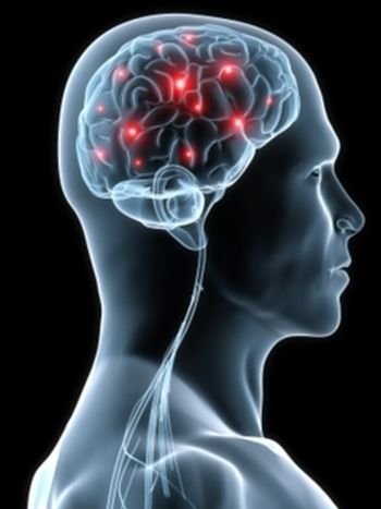
Perhaps it is not surprising those theories involving the solid tissues of the brain were difficult to conceive – after all, the brain’s solid substance has no visible moving parts. By the 17th century, however, the grip of humoral theory was weakening, in part due to the works of a new generation of anatomists who were describing the internal structure of the brain with increasing accuracy. Notably, the Englishman Thomas Willis (1621-1695), who coined the term ‘neurology’, argued that solid cerebral tissue has important functions. He still held that fluid-flow was the key to understanding brain function, but his focus was on the solid cerebral tissues and he showed that nervous function depends on the flow of blood to them. Today’s functional brain imaging technique (fMRI) shows that small local increases in blood flow are associated with the activation of nerve cells. That there is in effect a local ‘blushing’ of the brain when the neurons in that region are active is an observation that Willis might well have expected and enjoyed.
Among the more obvious problems of vital fluid and hydraulic models of nervous system function, and no doubt known to Willis, is that nerves are not hollow conduits. And even if they were, the speed of fluid movement through them could hardly be sufficiently swift for the rapidity with which sensations and motor commands seem to be conveyed by nerves. These and other inconsistencies with fluid models of the nervous system must have worried physicians of the stature of Willis and caused them pause for thought. But Willis remained a fluid theorist and the beginning of the end for the fruitless elaboration of such theories did not come until the discoveries attributed to Luigi Galvani (1737-1798). In the late 18th century he discovered the importance of electricity to the operation of the nervous system. As electrical mechanisms were to provide the necessary speed, attention inevitably turned from fluid to electrical models. Ironically, the last gasp of the legacy of Hippocrates and Galen is to be found in the interpretation Galvani himself placed on his own experiments with ‘animal electricity’. Having demonstrated that he could control the contractions of a frog muscle by applying electrical currents to the muscle’s motor nerve, Galvani claimed to have discovered that animal nerves and muscle contain an electric fluid’. A decisive leap of understanding however was achieved when Galvani and his contemporary Alessandro Volta (1745-1827) crucially together linked electricity to the functions of the nervous systems.
What neither Galvani nor Volta could know however is that the From humors to cells externally applied electrical stimuli were activating biological processes causing high-speed electrical impulses to travel along nerves to muscles, resulting in their contraction. It was not until the middle of the 19th century that the ability of nerves and muscles to generate rapidly propagating electrical impulses was confirmed by the German physiologist Du Bois-Reymond (1818-1896). This was a major impetus to the study of the physical workings of the brain and set the stage for the modern scientific era, which was launched in a most spectacular way at the dawn of the 20th century by the recognition of the cellular nature of the brain’s tiny functional units – the neurons.
The true cellular nature of the brain and of its mental functions was first recognized by the father of modern neuroscience, the Spanish neuroanatomist Santiago Ramon y Cajal (1852-1934). Although his proposition that the brain is a cellular machine may today seem commonplace, in fact it was revolutionary. In the later 19th century, and indeed in the early years of the 20th century, most neuroanatomists believed that the brain was not composed of cells at all – in spite of a universal recognition that all other organs and tissues in our bodies were. What was it about the brain that made it so difficult to see its cellular composition under the microscope?
Part of the answer is that brain cells are quite unlike any other cells. The very term ‘cell’ implies uniformity; simple structures defined by clear boundaries.
In contrast neurons are hugely diverse in morphology. They have exceedingly fine and profusely branched processes ramifying from the cell’s body and intermingling among the branches of other neurons. The complexity and diversity of their physical appearance easily exceeds that of all other cell types found in any other part of the body. All of this contributed to a rather confusing picture which anatomists found difficult to reconcile with a simple cellular model of brain structure. When viewed through a microscope the brain appeared to consist of a hopelessly tangled morass (a reticulum), without the distinct cell-defining boundaries that are so evident in other tissues. It was therefore not surprising perhaps that cell theory, the idea that tissues are composed of cells, was thought not to apply to the brain and a radical alternative model was proposed. This came to be called the ‘reticular theory’ of brain anatomy – a surprisingly resilient interpretation that persisted well into the 20th century. The reticular theory was wrong, but that was not the only problem with it. Scientific theories are allowed to be wrong so long as they are helpful, but the reticular theory, which held that the brain contains no discrete components, was actually obstructive to scientific progress. Progress was hindered by the concept of a machine without discrete functional components because without them it is impossible to formulate a plausible mechanism to explain how the brain might work. Scientists were sure the brain machine must have components and, given the complexity of what the brain does, lots of them. But what were they, what did they look like, and what did they do? It was clear that to understand the brain science had to identify the functional components of the brain’s microscopic structure.
Towards the end of the 19th century, the Italian anatomist Camillo Golgi (1843-1926) developed a way of highlighting the morphology of very few neurons in any particular region of the brain. It was a staining method that fitted the bill because it allowed individual neurons to be viewed unobstructed by the tangled mass of branched processes of neighboring cells. It incorporated the chemistry of photographic processing and it revealed individual neurons as dark, silver-impregnated silhouettes. Paradoxically, the crucial feature of Golgi’s method was that it hardly ever worked! Just one in a thousand or so neurons were ever revealed and these were scattered more or less randomly throughout the brain tissue. It was precisely because of this uncertain aspect of the method that neurons could for the first time be seen in their entirety disentangled from their neighbors. Immediately it was apparent that there are discrete cells in the brain, but they are astonishing cells – unlike any others. They differed markedly from one another, in particular with respect to the complex patterns of their numerous branched processes. Golgi’s method was the key to a new set of scientifically testable ideas about how the brain works. The reticular theory was about to be replaced by a far more powerful one called the neuron doctrine, the idea that the brain is composed of discrete cellular components.
The neuron doctrine is rightly attributed to Ramon y Cajal who, with the help of Golgi’s new staining method, made two profound propositions. The first quite simply is that the neuron is a cell. You might think that this must have been self-evident to anyone who bothered to view a brain treated with Golgi’s method. After all, cells in the brain would be clearly visible and thus by the evidence of one’s own eyes the reticular theory must be wrong. Somewhat astonishingly, however, in spite of the images provided by his own technique, even Camillo Golgi remained a convinced reticularist. The second of Cajal’s propositions was brilliantly insightful: neurons are structurally polarized with respect to function. For the first time, the workings of the brain were explicitly associated with the functions of physical structures at a microscopic level. Cajal concluded that a neuron’s function must be concerned with the movement and processing of information in the brain. He could only guess about the form in which information might be encoded or how it might move from place to place. In a stroke of genius, however, he postulated that it would be sensible for the components of function to impose directionality on information flow (or streaming as he called it). So he proposed that information flows in one direction, from an input region to an output region. The neuron’s cell body and its shorter processes, known as dendrites, perform input functions. Information then travels along the longest extension from the cell body, known as the axon, to the output region – the terminals of the axon and its branches that contact the input dendrites and cell body of another neuron.
Cajal was fascinated by the differences between the brains of markedly different organisms: human, worms, snails, insects, and so on. He thought comparisons of their brains might be instructive precisely because vast differences exist between the behaviour and intellectual capabilities of different creatures. There is unquestionably an enormous gulf between human and insect intelligence, so it would be reasonable to suppose that a comparison of their brains would expose how structural components reflect intelligence. Surely, the human brain should contain ‘high performance’ components and the insect brain markedly less sophisticated ones. But the difference between insect and human neurons does not at all betray the gulf between insect and human intelligence. Insect neurons are as complex and display as much diversity as neurons in the human cortex. Cajal himself expressed considerable surprise at this: the quality of the psychic machine does not increase with the zoological hierarchy. It is as if we are attempting to equate the qualities of a great wall clock with those of a miniature watch. Brains of the most advanced insects (honey bees) have about one million neurons, snails about 20,000, and primitive worms (nematodes) about 300. Contrast these numbers with the hundred billion or so that are required for human levels of intelligence. But the individual neurons of simple organisms operate with the very same electrical and chemical signalling machinery found in today’s most advanced brains. Like it or not, the astonishing conclusion from comparative studies is that the evolution of our brains, capable of such extraordinary feats, did not require the evolution of ‘super neurons’. The basic cellular components of mental functions are pretty much the same in all animals, the humble and the human.
In 1906 Cajal shared the Nobel Prize for Physiology and Medicine with Golgi, ‘in recognition of their work on the structure of the nervous system’. This was the first time that the Nobel Prize had been shared between two laureates. The award was controversial ecause the two disagreed on a crucially important matter – Golgi remained convinced that Cajal was wrong to reject the reticular theory. It was of course Golgi who was wrong and fundamentally so. Other questions over Golgi’s interpretations raised serious doubts in the minds of some of the scientists advising the Nobel Council as to the appropriateness of his nomination for the prize. But whatever the merits of the case for a shared prize, 1906 marked the beginning of the modern era in the neurosciences and it was the first of a series of Nobel Prizes to be awarded to neuroscientists over subsequent decades.
Cajal could not have anticipated the extraordinary advances in brain science that were about to be made. His recognition of the neurons as polarized units of information transmission was a defining moment in neuroscience. But at the start of the 20th century many questions about precisely how and in what form neurons signal information in the brain remained unanswered. By the middle of the 20th century, neuroscience had become the fastest growing discipline in the history of scientific endeavor and by the end of that century a more or less complete understanding, in exquisite molecular detail, of how neurons generate electrical and chemical signals would be achieved.
In this very short history of man’s discovery of the workings of the brain I cannot avoid reference to the discredited pseudo-science of phrenology, a theory developed by the idiosyncratic Viennese physician Franz Joseph Gall (1758-1828). Gall believed that the brain is the organ of the mind but he went much further and postulated that different distinct faculties of the mind, innate attributes of personality, and intellectual ability, are located in different sites in the brain. Gall reasoned that different individuals will have these innate faculties and that the degree of their development would be reflected in the size of the surface region of the brain that housed that particular faculty. These ideas have a very modern ring to them, but Gall thought that the skull would take the shape of the brain’s relief and therefore that the bumps on the surface of the skull could be ‘read’ as an index of various psychological aptitudes.
The practice of phrenology grew and flourished in Europe and then in America from about 1820, becoming a popular fad in the latter part of the 19th century before effectively dying out early in the 20th century (though in fact the British Phrenological Society was not disbanded until 1967). Its demise in the early 20th century coincided with the rapid accumulation of real evidence for the principle that many discrete mental functions are highly and specifically localized to particular parts of the brain. Much of the evidence came as a consequence of the First World War in the form of the many unfortunate victims of gun-shot and shrapnel lesions to specific regions of the brain that produced reproducible disorders. More recently, functional brain imaging techniques such as fMRI have shown beyond doubt that different cognitive functions are indeed localized to specific parts of the brain. So while the exaggerated claim of phrenologists to be able to read the mind from the bumps on the head was refuted, their premise was vindicated.
Imaging the future of brain research
The first high definition imaging system, called Computed Axial Tomography (CAT scanning), was developed in the 1970s. It is an X-ray-based technology that was used, and still is, as a medical diagnostic tool to resolve the position of brain tumours in the brain for example. In the past 30 years more powerful imaging technologies have been developed that have the potential to associate different cognitive functions with different structures in the brain. These techniques include most notably Positron Emission Tomography and Magnetic Resonance Imaging. When PET is used to link function to structure, increases in local blood flow and glucose consumption associated with increased neuronal activity are measured. A radioactive isotope, of glucose for instance, is injected into the blood stream and the high-energy photons that fly off in exactly opposite directions from the site of an emitting isotope are detected by an array of detectors that encircle the head. The detectors facing one another on opposite sides of the head will simultaneously detect the two photons generated from the same place within the brain. By the integration of simultaneous photon detection in the array, the source of the isotope can be calculated. In this way a computer builds an image of the structures that contain the isotope. In other applications of PET, the radioactive label is attached to molecules that bind to particular receptors, thus revealing the distribution of neurotransmitter systems receptors in the brain, for example.
A more sensitive technique, importantly that does not involve radioactive tracers, is Magnetic Resonance Imaging or MRI. Briefly the technique involves the pulsing of a strong external magnetic field, which evokes transient magnetic responses within the brain. The evoked magnetic signals are used to compute 2D and 3D images of the brain’s structure. This technique can be used for purely structural studies, as it was in the experiments on London taxi drivers that showed they have a larger than expected hippocampus. But in its most interesting experimental application it provides images of the brain in action. When used to reveal active regions of the brain involved in particular functions, the technique is known as functional MRI, or fMRI for short. To understand how fMRI works, and to appreciate its limitations, it is important to realize that it does not image the electrical activity of neurons directly. It monitors the indirect consequence of their activity. When a region of the brain is actively working, more neurons in that region will require more glucose and oxygen. This is a consequence of two interesting facts. First, it seems neurons only store enough energy for the briefest of bouts of activity. If neurons are active long enough, they need refueling to enable them to produce the energy storage molecule ATP required to recharge their batteries. An active brain region therefore may have a significantly higher metabolic demand for oxygen and glucose than a quiescent region. A simple solution to this problem would be to pump more blood into the active brain, much in the same way that a muscle is supplied with more blood when exercised vigorously. However unlike a muscle, which becomes engorged with blood and swells when exercised, the brain is confined by the skull and cannot be allowed to swell significantly. The solution to this tricky problem is to maintain a constant overall blood-volume in the brain and to arrange for blood to be diverted preferentially to active regions. Blood is diverted by the ability of blood vessels in the brain to dilate in response to signals coming from nearby active neurons. Dilation reduces resistance to blood flow, thereby increasing the supply of blood to the region of elevated neuronal activity. We are not really sure how the blood vessels ‘know’ that nearby neurons are hyperactive. It is likely however that the signal for blood vessel dilation is the gas nitric oxide (NO), because NO causes the relaxation of muscle cells in the walls of blood vessels. It is thought that NO-producing neurons sense increased activity of nearby neurons and respond by producing NO in the same region – thus coupling increased neural activity to increased blood flow in that region. In detecting regions of increased blood flow, fMRI recognizes the different magnetic signatures of oxygenated and deoxygenated hemoglobin. When neurons in a brain region are sufficiently active for long enough, blood in their vicinity becomes oxygen depleted. This is followed by an increased flow of oxygenated blood to that region; quite literally there is a local blushing of the brain. The fMRI technique is responsive to the blushing and indirectly assigns increased neural activity to that region at a spatial resolution of just a few cubic millimeters. It is in this way that we now have a far more fine-grained functional map of the brain than was previously possible. Bold claims are now being made about complex cognitive functions: where in the brain we recognize faces and words, where executive functions are carried out, where false memories are located, and so on.


